Vocabulary
|
|
|
|---|
Let's Recall!

Case Studies
Do you remember what a case study is from unit 1? Let's go over it once more!
A case-study is an in-depth study of an individual or a small group. Usually, case studies are done on people with rare circumstances.
A case-study was done on Phineas Gage, who had an iron pole penetrated through his skull and brain🧠 No one else ever had this issue/injury, but scientists did an in-depth study on him to advance their understanding of brain function.
Split-Brain
Case studies could also be done on split-brain patients. In the past, a surgery may have been done to eliminate seizures in which doctors would severe the corpus callosum (which connects the two brain hemispheres).
Doing case studies on split-brain patients helped scientists gain a deeper understanding of how the brain works and what each part of the brain is responsible for. Interestingly enough, they found out that each hand✋ could be drawing a completely different image at the same time without messing up. It's really cool to see this happen!
Roger Sperry was one of the brain neuroscientists that advanced research with split-brain patients. He was able to gain insight about each hemisphere of the brain:
- Left Hemisphere - Controls right hand, spoken language, written language✍, mathematical and logical thought processes, analysis, and reading📚
- Right Hemisphere - Controls left hand, nonverbal (visual) perception. Is responsible for musical🎵 and artistic processing🎨 and emotional thought- Think: Left Deep Thinking, Right Creative Mind
Examining Brain Structures and Function
Advancements in technology have allowed psychologists to study the brain in new and exciting ways. Lesioning may be used in circumstances to destroy certain, selective, parts of the brain in order to reduce a behavior. We can also stimulate areas of the brain to generate a response. For example, I could electrically stimulate your motor cortex to make you raise your hand✋
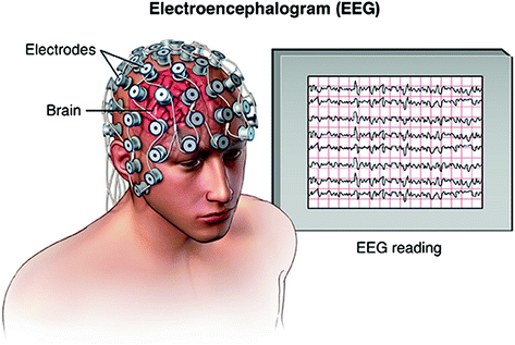
Electroencephalograms (EEGs) and Positron Emission Tomography (PET) scans are both ways in which we can see the activity of the brain. EEGs measure and record brain waves in various states, such as dreaming and sleeping, to study cognition. PETs use glucose to monitor which parts of the brain light up when patients are given various tasks and produces color graphics. Using PETs, you could visualize changes in the brain as it functions. This is because when a neuron is active, blood flow in increased and more glucose and oxygen is brought into the brain. When using PET, radioactively tagged glucose is injected into the bloodstream, and this is detected through a PET scanner. It then shows where the glucose is going when and at which instants.
Computer Tomography (CT) scans and Magnetic Resonance Imaging (MRI) create visual images of the structure of the brain. CT scans use a variety of X-ray photographs to create the image of a two-dimensional slice. MRIs use various magnetic fields and radio waves to create an image of the brain’s soft tissue.
fMRIs are unique in that they can show us both the physical structure of the brain as well as activity and function. This has allowed us to gain more insight into which parts of the brain are responsible for specific tasks and abilities.
Summary:
| Technology | Uses | Benefits | Limitations |
|---|---|---|---|
| CT (CAT) | Two-dimensional image of brain using X-rays | Shows structure of brain and any damages | Does not show function of brain |
| PET | Radioactive glucose tracked down to show metabolism by the brain | Records brain activity | Less precise than fMRI and exposure to radiation |
| EEG | Electrodes placed on head and graphical image is produced | Useful with sleep and epilepsy research | No structure or function of brain |
| fMRI | Measures change in blood flow and creates 3D image | More precise than PET scan with functional picture of brain | Brain areas activate for different reasons but unable to detect this |
🎥Watch: AP Psychology - Tools of Discovery for the Brain